.png)
Physical Therapist Resource Hub
Clinical Decision Support in Hip Preservation
Physical therapists are often the first clinicians to evaluate young athletes with hip pain. Determining when to continue structured rehabilitation, when advanced imaging is appropriate, and when referral for hip preservation evaluation is indicated can be complex — particularly in cases involving femoroacetabular impingement (FAI), labral pathology, dysplasia, or hip microinstability.
This resource hub is designed as a clinical decision support framework. It provides practical algorithms, differentiation tables, imaging interpretation guidance, and referral indicators grounded in an indications-first philosophy.
The goal is not to accelerate surgical referral.
The goal is to support appropriate decision-making based on patient trajectory, functional response, and structural stability.
Core Principle:
Imaging supports clinical decision-making — it does not replace it.
Morphology alone does not determine intervention.
Functional limitation, symptom reproducibility, mechanical instability, and failure of structured rehabilitation remain the primary drivers of referral consideration.
Stepwise Clinical Algorithm
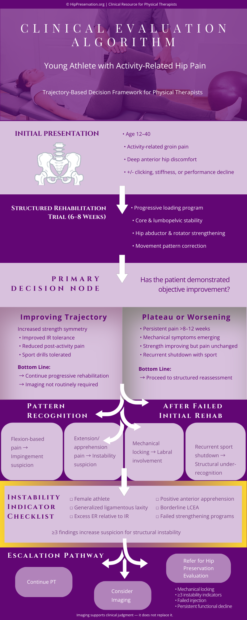

Clinical Interpretation: Understanding the Gray Zone
Most young athletes with hip pain do not fall cleanly into “rehab success” or “surgical candidate” categories. The majority exist in a diagnostic gray zone characterized by partial strength improvement, persistent activity-related pain, and imaging findings that may or may not correlate with symptoms.
Clinical trajectory — not isolated morphology — determines progression toward referral.

The majority of patients fall within the “Monitor / Reassess” column. Referral becomes progressively weighted when symptoms persist beyond 12 weeks despite structured rehabilitation, mechanical symptoms increase in frequency, or instability indicators accumulate. Escalation decisions should reflect a pattern of failed trajectory rather than a single exam finding.
Escalation is driven by failure of progression — not the presence of morphology alone.
.png)
Misclassification between impingement-dominant and instability-dominant pathology is one of the most common contributors to persistent postoperative pain and failed conservative management. While cam morphology is common in asymptomatic athletes, instability — particularly borderline dysplasia and capsular laxity — is frequently under-recognized.
Careful attention to rotational patterns, apprehension signs, and symptom reproduction arc improves diagnostic accuracy.
Clinical Takeaway: Instability is often under-recognized in the young athletic population.
Imaging Interpretation: Context Over Morphology
Imaging findings are common in asymptomatic athletic populations. Labral tears, cam morphology, and even mild dysplasia may be present in patients without functional limitation. For this reason, imaging must support clinical findings — not dictate escalation independently.
The relevance of imaging is determined by symptom reproduction, mechanical instability, and failure of structured rehabilitation.
Labral Tears on MRI
Labral tears are frequently identified on MRI in athletes without symptoms. The presence of a tear alone does not establish surgical indication. Mechanical locking, reproducible catching, and persistent activity limitation are stronger clinical drivers than imaging alone.


Cam Morphology and Alpha Angle
Cam morphology is prevalent in high-level athletes, particularly males participating in cutting and pivoting sports. Increased alpha angle does not inherently require intervention. Clinical correlation with flexion-based pain and functional limitation is required before escalation.
Dysplasia and the Limits of LCEA Alone
Borderline dysplasia cannot be diagnosed by lateral center-edge angle in isolation. Assessment must include anterior coverage, acetabular version, Tönnis angle, and clinical instability signs. Under-recognition of instability is a common contributor to failed arthroscopy.

Imaging supports clinical decision-making.
It does not replace functional assessment and trajectory analysis.
Diagnostic Injection: What the Response Actually Tells You
Image-guided intra-articular hip injections are frequently used in the evaluation of persistent hip pain. However, interpretation of injection response requires nuance. Relief of pain does not automatically imply surgical indication, and lack of response does not definitively exclude intra-articular pathology.
Injection results must be interpreted in the context of mechanical symptoms, instability signs, and functional limitation.

Complete symptom relief confirms an intra-articular pain source but does not determine the optimal treatment pathway. Persistent mechanical symptoms or functional shutdown after adequate rehabilitation increase referral weight. Conversely, absent relief should prompt reconsideration of extra-articular contributors, lumbopelvic mechanics, or subtle instability patterns.
Injection response clarifies pain origin — it does not independently determine intervention.
Patterns of Failed Arthroscopy
Why Some Scopes Do Not Fully Resolve Symptoms
Hip arthroscopy can be highly effective in appropriately selected patients. However, persistent or recurrent symptoms following arthroscopy are not uncommon, particularly when underlying structural instability or version abnormalities were not fully recognized preoperatively.
Understanding the common patterns associated with suboptimal outcomes helps physical therapists identify patients who may benefit from further structural evaluation rather than repeated rehabilitation alone.
Unrecognized Structural Instability
One of the most frequent contributors to persistent postoperative pain is under-recognized instability. Borderline dysplasia, capsular laxity, and version abnormalities may not be fully appreciated on initial evaluation.
Patients may initially improve, only to experience recurrent pain with return to sport.
Clinical Clues:
-
Female athlete
-
Persistent apprehension
-
Excess external rotation
-
Pain with extension-based activity
Instability misclassified as isolated impingement can limit arthroscopic success.
Capsular Insufficiency
Excessive capsulotomy or inadequate capsular repair may contribute to persistent microinstability. These patients often describe vague discomfort, fatigue pain, or loss of confidence in dynamic activities.
Clinical Pattern:
-
Deep ache rather than sharp mechanical pain
-
Difficulty with pivoting sports
-
Apprehension without true locking
Version Abnormalities
Femoral or acetabular version abnormalities can alter joint biomechanics and may not be fully corrected during arthroscopy. Persistent rotational discomfort or asymmetrical motion may remain.
Clinical Clues:
-
Persistent rotational pain
-
Asymmetry not explained by strength deficits
-
Limited improvement despite good rehab adherence
Incomplete Cam Resection
In some cases, residual impingement morphology may persist.
Clinical Pattern:
-
Flexion-based pain remains primary complaint
-
FADIR remains strongly positive
-
Mechanical symptoms continue
This is less common in experienced centers but remains a recognized contributor.
Non-Intra-Articular Contributors
Not all persistent pain after arthroscopy is structural. Extra-articular sources such as:
-
Iliopsoas irritation
-
Core weakness
-
Abductor insufficiency
-
Lumbopelvic dysfunction
must also be considered.
Rehabilitation remains critical in these cases.
Clinical Perspective
Persistent symptoms following hip arthroscopy are rarely the result of a single variable. Careful reassessment of structural stability, version alignment, and rehabilitation progression is essential before assuming surgical failure. Collaborative discussion can help determine whether continued rehabilitation, imaging reassessment, or structural evaluation is most appropriate.
Not all persistent pain after arthroscopy indicates surgical error.
Many cases reflect unrecognized instability, version complexity, or multifactorial contributors.

Return-to-Sport Risk Stratification Framework
Clinical Markers of Readiness vs Risk
Return to sport following hip rehabilitation or hip arthroscopy requires more than strength symmetry alone. Persistent instability patterns, fatigue pain, and mechanical symptoms may increase reinjury risk even when objective metrics appear adequate.
A structured risk stratification model helps guide progression decisions and identify patients who may benefit from further evaluation.
Low Risk Return to Sport

-
Symmetric hip strength (within 10%)
-
Pain-free sport-specific drills
-
No apprehension with extension/rotation
-
No mechanical locking or catching
-
Full tolerance to progressive loading
Patients meeting these criteria typically progress safely with structured return-to-play protocols.
.png)
Caution Zone
-
Mild fatigue-related groin pain
-
Slight asymmetry in internal rotation
-
Apprehension with high-speed pivoting
-
Inconsistent tolerance to volume increases
-
Fear avoidance behaviors
These athletes may require load modification, neuromuscular retraining, and closer monitoring before full clearance.

Elevated Risk
-
Recurrent mechanical locking
-
Extension-based instability pain
-
Persistent apprehension sign
-
Progressive loss of ROM
-
Shutdown during sport participation
Recurrent shutdown or mechanical symptoms warrant structural reassessment prior to unrestricted return.
Clinical Integration
Return-to-sport decisions should integrate mechanical stability, symptom reproducibility, and fatigue response — not strength testing alone. In cases of repeated sport intolerance despite adequate rehabilitation, structural evaluation may be appropriate.
Recurrent shutdown is not deconditioning. It is a diagnostic signal.
Collegial Case Collaboration & Educational Resources
Collaborative Case Discussion
Hip preservation decision-making often requires integration of rehabilitation trajectory, imaging findings, and structural stability assessment. In cases that fall within the clinical gray zone, direct discussion can help clarify whether continued rehabilitation, further imaging, or structural evaluation is most appropriate.
Collegial case review is welcomed for complex or uncertain presentations.
What to Include in a Case Summary
-
Patient age and activity level
-
Duration of symptoms
-
Rehabilitation timeline and response
-
Mechanical symptoms (if present)
-
Imaging summary (if available)
-
Injection response (if performed)
For case discussion:
Case discussions are intended to support collaborative care and are not a substitute for formal medical consultation.
Educational Resource for Physical Therapists
For physical therapists interested in a broader framework of hip preservation principles — including early diagnosis, conservative management, surgical indications, and post-operative rehabilitation collaboration — the following presentation may be helpful.
